
The ventricles are more richly innervated by sympathetic fibers than parasympathetic fibers.
#High heart rate plus#
The cardioaccelerator center also sends additional fibers, forming the cardiac nerves via sympathetic ganglia (the cervical ganglia plus superior thoracic ganglia T1–T4) to both the SA and AV nodes, plus additional fibers to the atria and ventricles. īoth sympathetic and parasympathetic stimuli flow through the paired cardiac plexus near the base of the heart. Normally, vagal stimulation predominates as, left unregulated, the SA node would initiate a sinus rhythm of approximately 100 bpm. This is a similar concept to tone in skeletal muscles. During rest, both centers provide slight stimulation to the heart, contributing to autonomic tone. The cardioaccelerator regions stimulate activity via sympathetic stimulation of the cardioaccelerator nerves, and the cardioinhibitory centers decrease heart activity via parasympathetic stimulation as one component of the vagus nerve. Nervous influence over the heart rate is centralized within the two paired cardiovascular centres of the medulla oblongata.

It is also influenced by central factors through sympathetic and parasympathetic nerves. The heart rate is rhythmically generated by the sinoatrial node. Influences from the central nervous system Cardiovascular centres This section discusses target heart rates for healthy persons, which would be inappropriately high for most persons with coronary artery disease. Most involve stimulant-like endorphins and hormones being released in the brain, some of which are those that are 'forced'/'enticed' out by the ingestion and processing of drugs such as cocaine or atropine. There are many ways in which the heart rate speeds up or slows down.

However, heart rates from 50 to 60 bpm are common among healthy people and do not necessarily require special attention. Bradycardia is defined as a resting heart rate below 60 bpm. Normal resting heart rates range from 60 to 100 bpm. Īs water and blood are incompressible fluids, one of the physiological ways to deliver more blood to an organ is to increase heart rate. Therefore, stimulation of the accelerans nerve increases heart rate, while stimulation of the vagus nerve decreases it. The accelerans nerve provides sympathetic input to the heart by releasing norepinephrine onto the cells of the sinoatrial node (SA node), and the vagus nerve provides parasympathetic input to the heart by releasing acetylcholine onto sinoatrial node cells. While heart rhythm is regulated entirely by the sinoatrial node under normal conditions, heart rate is regulated by sympathetic and parasympathetic input to the sinoatrial node. Problems playing this file? See media help. Abnormalities of heart rate sometimes indicate disease. When the heart is not beating in a regular pattern, this is referred to as an arrhythmia. When a human sleeps, a heartbeat with rates around 40–50 bpm is common and is considered normal.

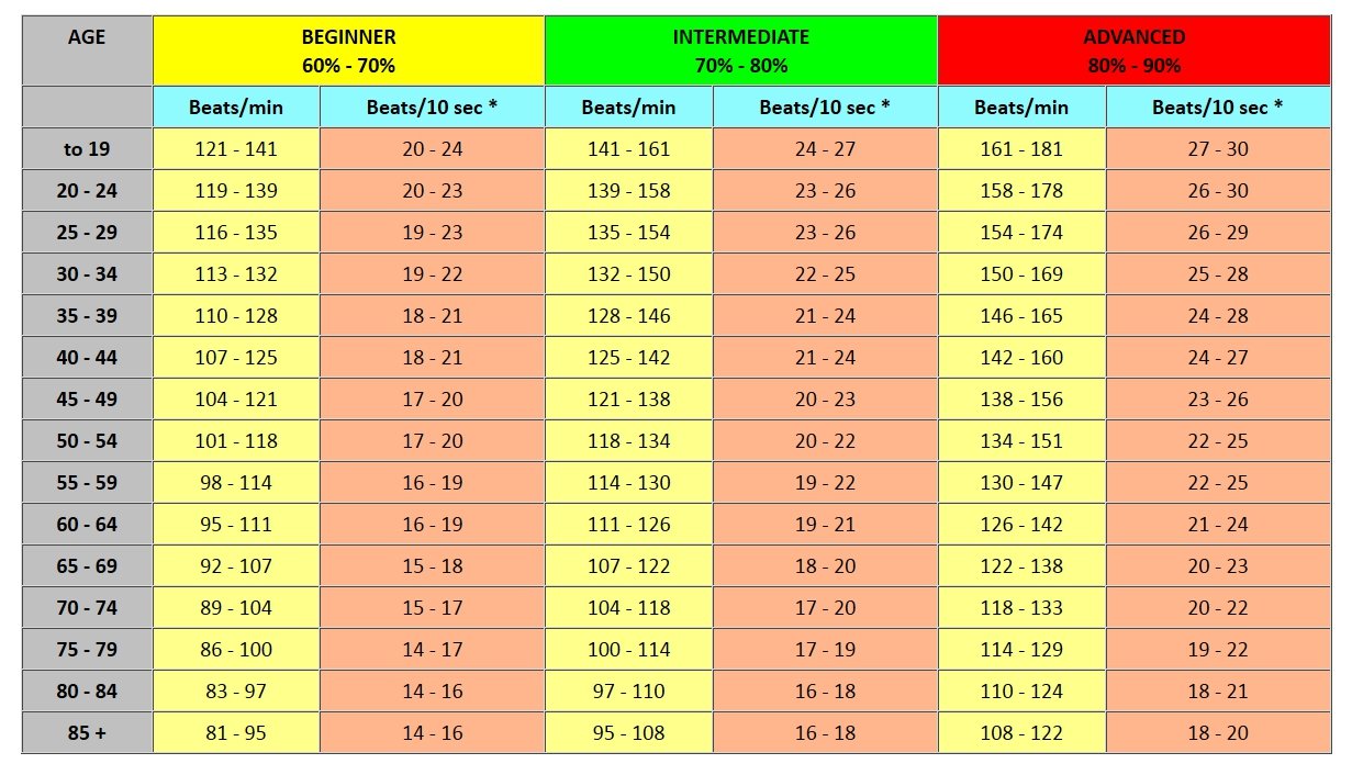
Bradycardia is a low heart rate, defined as below 60 bpm at rest. Tachycardia is a high heart rate, defined as above 100 bpm at rest. The American Heart Association states the normal resting adult human heart rate is 60-100 bpm. It is usually equal or close to the pulse measured at any peripheral point. The heart rate can vary according to the body's physical needs, including the need to absorb oxygen and excrete carbon dioxide, but is also modulated by numerous factors, including (but not limited to) genetics, physical fitness, stress or psychological status, diet, drugs, hormonal status, environment, and disease/illness as well as the interaction between and among these factors. Heart rate (or pulse rate) is the frequency of the heartbeat measured by the number of contractions of the heart per minute ( beats per minute, or bpm). Speed of the heartbeat, measured in beats per minute


 0 kommentar(er)
